I wish to create this animation from the perspective of the surgeon, as though it is the viewer’s hands performing the procedure. The environment will be modern, sterile and bright as it is in an operating room. The materials will be metals (the OR table and all of the surgical instruments), rubber (the surgeon’s gloves) and cloth (drapery and surgeon’s scrubs) with subsurface scattering material for skin, dielectrics for the blood, and materials yet to be determined for bone and muscle (something glossy and wet looking).
I would very much like to create something with clean models; I do not want to create an incredibly bloody scene with spatter and shredded muscle, but rather a clean scene with minimal blood and text-book clean muscles in such a way that the anatomy being manipulated in the animation can easily be identified by the viewer. I am going for a realistic but clean look. In a perfect world I would like to focus not only on creating beautiful textures for this animation but also on the realism of the physics behind cutting and manipulating different tissues. With time constraints I have chosen to focus more heavily on creating realistic physics, and hope to have the time to infuse a bit of good texturing. I will stick with Maya to do all of my modeling, as I have found good tutorials for foot, leg and hand modeling. Obviously these models will need to be rigged, and Maya Muscle will be used to help create the realism behind the tissue manipulation. If necessary, motion capture may be used to help animate the fine motor movements of the hand during certain surgical manipulations. The surgical instruments will be modeled in Maya as well, and nCloth will be used to simulate the drapery and to clothe the surgeon’s hands in latex gloves.
As is appropriate for any kind of instructional video, narration will be necessary. Also, as the actual operation takes a total of thirty minutes to an hour, steps will be shortened and spliced together in After Effects in order to make this animation the appropriate length.
The following is exactly what would need to be animated in order to depict a below knee amputation: The procedure will start with a physical examination of the uncovered, affected leg to be operated on. A cloth will be placed around the gangrenous area. The surgeon will move on to drawing incision marks with a marker along the sides and circumference of the leg. Then, the actual incision is performed along these marks with a scalpel. An electrocautery device is used to cut through the anterior and lateral muscles. An anterior vascular bundle is ligated to prevent bleeding. Then scrapers and retractors are used to pull back tissue from the tibia and fibula. A reciprocating saw is used to separate the tibia and a bone cutter is used to section the fibula. The only remaining tissue attaching the leg to the body, at this point, are posterior muscles that are quickly sliced through with a scalpel in a downward, curved fashion. The gangrenous leg is at this point completely detached from the body and is quickly removed from the operating area. The sectioned arteries poking out of the freshly sliced posterior muscles are quickly clamped to prevent bleeding out, and then the posterior tibial nerve is divided via the Guilltine technique, causing the nerve stump to retract within the muscle mass. In the same manner, the sural nerve is divided. The surgeon bevels the end of the tibia with the reciprocating saw and then rounds the bone carefully with a fine rasp. The hanging posterior skin and muscle flap is tailored with a scalpel to remove excess muscle that would get in the way of closing the flap. The surgeon then lifts the flap up and around to meet the front of the leg. Holding the flap in this closed position, the surgeon staples the wound closed and then supplements this closure with stitches. The operation at this point is complete and this would be the end of the animation.
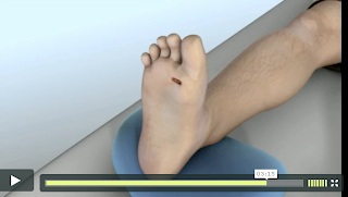
1. Reference from XVIVO Scientific Animation. I would love to accomplish this level of realism in the modeling of the foot and leg.
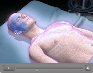
2. Reference from Argosy Medical Animation. I like the sterile environment and the subtle use of cloth folding, as well as the surgical basins in the background detail.
3. Reference from Radius Medical Animation. The use of extreme close-ups such as this is something I’d like to incorporate. Unfortunately, due to time constraints, it may be difficult to render out an entire animation with the use of depth of field, something I would really prefer to be able to do.


No comments:
Post a Comment